Picture Of Forearm Tendons - sinew - Wiktionary / Choose from up to 5 unique, high quality paper types to meet your creative or business needs.
Picture Of Forearm Tendons - sinew - Wiktionary / Choose from up to 5 unique, high quality paper types to meet your creative or business needs.. Human anatomy for the artist: Symptoms of forearm tendinitis include pain along the forearm, tenderness, and stiffness. We can tell this is a ventral view of the forearm because we can see the palmar aponeurosis (a thin, tendinous sheath that is only on the palmar side of the hand) and. The common extensor tendon is a soft tendon that's located in the forearm. Knowledge of normal anatomy and variations in the tendons of extensor muscles is important for identification of accessory.
We can tell this is a ventral view of the forearm because we can see the palmar aponeurosis (a thin, tendinous sheath that is only on the palmar side of the hand) and. It's most commonly caused by. Your walls are a reflection of your personality, so let them speak with your favorite quotes, art, or designs printed on our custom posters! Today is your day to take your inspired action before the full picture is in clear view! Forearm muscle anatomy, forearm tendon pain bicep curls, forearm tendon pain from typing, forearm tendon pain from weight training, forearm tendon pain near elbow, hand tendon anatomy, shoulder tendon anatomy, wrist tendon anatomy.
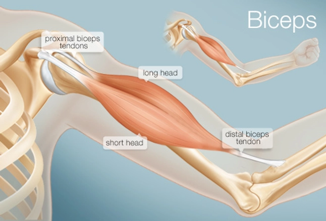
The Biceps (Human Anatomy): Function, Diagram, Conditions ... from img.webmd.com Forearm muscle anatomy, forearm tendon pain bicep curls, forearm tendon pain from typing, forearm tendon pain from weight training, forearm tendon pain near elbow, hand tendon anatomy, shoulder tendon anatomy, wrist tendon anatomy. We can tell this is a ventral view of the forearm because we can see the palmar aponeurosis (a thin, tendinous sheath that is only on the palmar side of the hand) and. Injuries the common conditions within the tendons throughout the elbow joint comprising of the tennis elbow, and the golfer's elbow, which occur from an overuse injury to the tendons or result from. The tendons of extensor muscles help stabilize the hand during forced graping and provide the loosness needed for sensitive finger movements independent from each other. Today is your day to take your inspired action before the full picture is in clear view! They are shown in the illustration below. Originates from the anterior surface of the ulna and attaches to the anterior surface of the radius. Knowledge of normal anatomy and variations in the tendons of extensor muscles is important for identification of accessory.
Originates from the anterior surface of the ulna and attaches to the anterior surface of the radius.
Forearm flexor muscles, labeled drawing. Today is your day to take your inspired action before the full picture is in clear view! The tendons of extensor muscles help stabilize the hand during forced graping and provide the loosness needed for sensitive finger movements independent from each other. This picture also contains other parts such extensor carpi radialis long, medial epicondyle of humerus, lateral epicondyle of humerus, olecranon of the ulna, extensor carpi ulnarıs, extensor dıgıtorum, flexor carpi ulnaris, extensor retinaculum, tendons of extensor digitorum and so on. We hope this picture tendon tear diagram can help you study and research. Tendinitis is an inflammation or swelling of a tendon. Webmd's achilles tendon anatomy page provides a detailed image and description of its function as well as conditions that affect the achilles tendon. 12 photos of the forearm tendon anatomy picture. The muscle of the common extensor tendon that is nearest this side of the arm is the extensor carpi ulnaris, which attaches to the proximal end of the fifth metacarpal, or the palm bone beneath the pinky. No tension in these tendons tolerated at all. It's most commonly caused by. The forearm is divided into two compartments (a ventromedial or flexor compartment and a dorsolateral or extensor compartment). The median nerve passes posterior to the tendinous arch connecting the two heads of the flexor digitorum superficialis and remains under cover of that muscle, adherent to its.
Forearm pain from muscle or tendon injuries can be quite debilitating. Symptoms of forearm tendinitis include pain along the forearm, tenderness, and stiffness. Choose from up to 5 unique, high quality paper types to meet your creative or business needs. How to treat forearm tendonitis. The picture above is an example of a great stretch for the inner forearm muscles and tendons, do this stretch before during and after you climb both indoor and outdoor.
Picture tests in anatomy arm and forearm 2 - YouTube from i.ytimg.com I figure that at this rate, i'm probably a minimum of two months out from being able to safely climb at the level i was at again. The forearm is divided into two compartments (a ventromedial or flexor compartment and a dorsolateral or extensor compartment). Median nerve (anterior interosseous branch). Tendons are the connective tissues that connect muscle to bone. These pictures of this page are about:forearm tendons and ligaments. Human anatomy for the artist: The median nerve passes posterior to the tendinous arch connecting the two heads of the flexor digitorum superficialis and remains under cover of that muscle, adherent to its. The term forearm is used in anatomy to distinguish it from the arm.
It hurts, not just when you lift or exercise, but also when you do everyday tasks, even something as basic as typing or moving the mouse on your computer.
Lesson on the anatomy of the forearm: Its muscle belly is in the forearm and then travels along the inside of the forearm and. Tendons are fibrous cords, similar to a rope, and are made of collagen. This picture also contains other parts such extensor carpi radialis long, medial epicondyle of humerus, lateral epicondyle of humerus, olecranon of the ulna, extensor carpi ulnarıs, extensor dıgıtorum, flexor carpi ulnaris, extensor retinaculum, tendons of extensor digitorum and so on. They are shown in the illustration below. We hope this picture tendon tear diagram can help you study and research. Read about ruptured tendon symptoms, treatment, and prognosis, whether it's an achilles tendon rupture or the tendon rupture is in the quadriceps, finger, ankle, hand, wrist, elbow, shoulder, knee, or anywhere else in the. 12 photos of the forearm tendon anatomy picture. Posted by health life media team on june 17, 2017. Knowledge of normal anatomy and variations in the tendons of extensor muscles is important for identification of accessory. The forearm is the region of the upper limb between the elbow and the wrist. Anatomy diagrams of shoulder, arm, elbow, forearm, wrist and hand. The picture above is an example of a great stretch for the inner forearm muscles and tendons, do this stretch before during and after you climb both indoor and outdoor.
A tendon is the fibrous tissue that attaches muscle to bone in the human body. Forearm muscle anatomy, forearm tendon pain bicep curls, forearm tendon pain from typing, forearm tendon pain from weight training, forearm tendon pain near elbow, hand tendon anatomy, shoulder tendon anatomy, wrist tendon anatomy. Picture of the achilles tendon. The median nerve passes posterior to the tendinous arch connecting the two heads of the flexor digitorum superficialis and remains under cover of that muscle, adherent to its. Originates from the anterior surface of the ulna and attaches to the anterior surface of the radius.
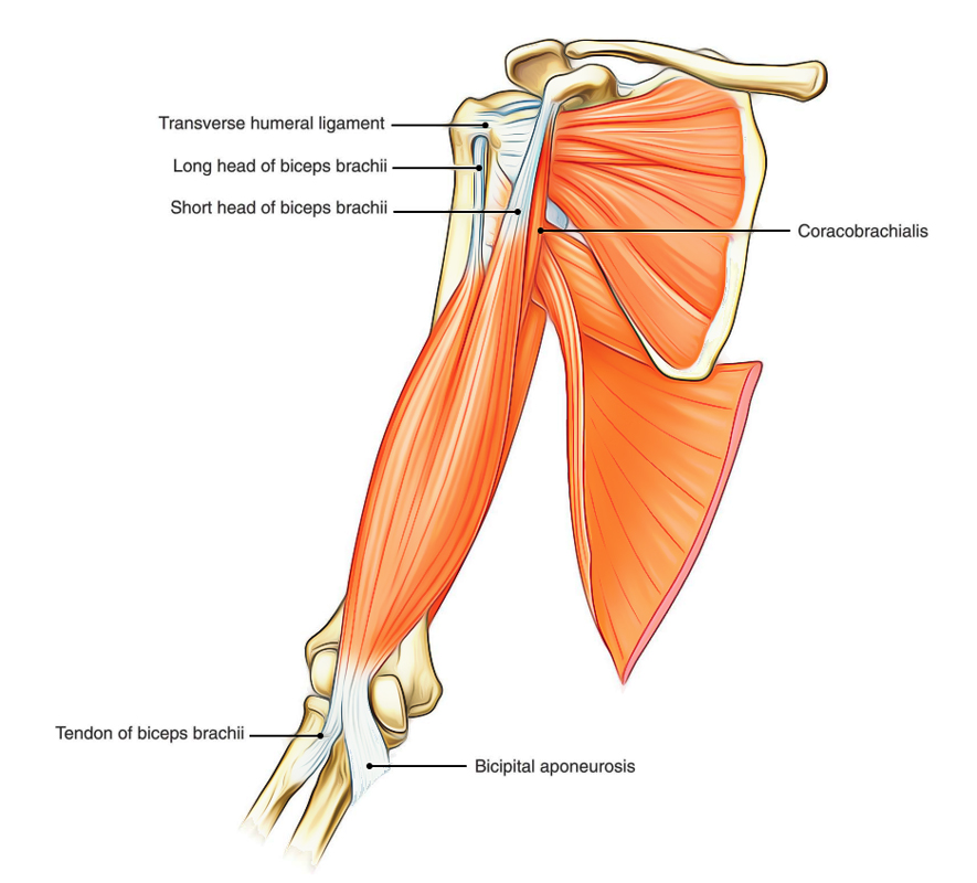
Easy Notes On 【Muscles of the Upper Arm】Learn in Just 3 ... from www.earthslab.com The forearm is divided into two compartments (a ventromedial or flexor compartment and a dorsolateral or extensor compartment). The tendons of extensor muscles help stabilize the hand during forced graping and provide the loosness needed for sensitive finger movements independent from each other. Webmd's achilles tendon anatomy page provides a detailed image and description of its function as well as conditions that affect the achilles tendon. These pictures of this page are about:forearm tendons and ligaments. Posted by health life media team on june 17, 2017. Choose from up to 5 unique, high quality paper types to meet your creative or business needs. Also within a half an hour after any climbing make sure you have eat some sort of protein, i don't have scientific number saying how much. The muscle of the common extensor tendon that is nearest this side of the arm is the extensor carpi ulnaris, which attaches to the proximal end of the fifth metacarpal, or the palm bone beneath the pinky.
Your walls are a reflection of your personality, so let them speak with your favorite quotes, art, or designs printed on our custom posters!
Webmd's achilles tendon anatomy page provides a detailed image and description of its function as well as conditions that affect the achilles tendon. Human hand tendon diagram (page 1) hand tendons diagram muscle blank drawing these. It hurts, not just when you lift or exercise, but also when you do everyday tasks, even something as basic as typing or moving the mouse on your computer. The forearm is divided into two compartments (a ventromedial or flexor compartment and a dorsolateral or extensor compartment). No tension in these tendons tolerated at all. Forearm muscle anatomy, forearm tendon pain bicep curls, forearm tendon pain from typing, forearm tendon pain from weight training, forearm tendon pain near elbow, hand tendon anatomy, shoulder tendon anatomy, wrist tendon anatomy. In most cases, conservative treatments such as avoiding any activity that. Knowledge of normal anatomy and variations in the tendons of extensor muscles is important for identification of accessory. Pain, swelling, and redness of the forearm are the most commonsymptoms of the condition. Those two tendons come from the palmaris longus muscle and the flexor carpi radialis muscle. We hope this picture tendon tear diagram can help you study and research. These pictures of this page are about:forearm tendons and ligaments. The two most common types of tendinitis are on the inside or outside of your elbow.